Türkçe
 English

Zirconium (zirconium dioxide), developed to eliminate the aesthetic and biological disadvantages created by metal-supported porcelains in prosthetic dentistry, is a restoration material we frequently use in our clinical practice. This material, which shows high biocompatibility with tissues, has become one of our main options in restorative procedures because it offers high resistance to chewing forces and can mimic natural tooth enamel with its optical properties.
Zirconium is used in dentistry in the form of its chemically stabilized crystalline structure (Y-TZP - yttrium-stabilized tetragonal zirconia polycrystal). This special structure provides a phase transformation (strengthening mechanism) that stops the progression of microcracks that may form within the material.
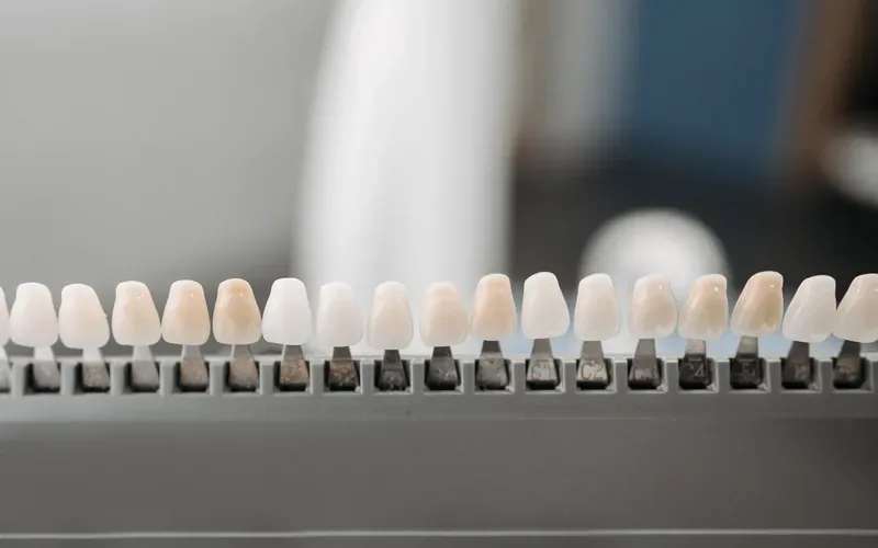
The gray reflection (metal corrosion or marginal discoloration) observed over time at the gum margin in traditional metal-based crowns is not seen in zirconium restorations. Light transmission (translucency) can be calibrated to match the color and opacity of the patient's natural teeth; this provides high aesthetic integration, especially in anterior (front) restorations. In addition, because microbial dental plaque retention (adhesion) on the zirconium surface is very low, it directly contributes to the preservation of gum health (periodontal tissues) and carries no risk of allergic reactions.
As a physician, when deciding on a zirconium crown, we analyze the amount of tissue loss in the patient's tooth structure, periodontal status, and occlusal (chewing) dynamics as a whole. The main application areas are:
Teeth with extensive tissue loss that cannot be restored with a composite filling due to decay, fracture, or trauma sequelae.
Full crown restoration of teeth that have undergone root canal treatment (endodontic treatment) and have become more fragile because their structural integrity and flexibility have decreased.
Implant-supported fixed prostheses used in the treatment of missing teeth (both single-unit crowns and bridges).
Aesthetic prosthetic corrections planned as an alternative to orthodontic treatment in cases of diastema (gapped teeth) or mild crowding.
The procedure usually begins under local anesthesia with the tooth in question being reduced circumferentially by an average of 1 - 1.5 mm from the enamel and dentin tissues (preparation). Clearly defining the gum margins and making a step-type cut of the "chamfer" or "shoulder" type is the most critical stage to ensure marginal adaptation and avoid violating the biological width.
After preparation, the impression stage begins. In current clinical practice, instead of traditional impression materials, three-dimensional optical records are mostly taken using intraoral scanners. This digital data is transferred to computer-aided design (CAD) software, and the anatomical form of the tooth is designed with millimetric precision. After the design is approved, computer-aided manufacturing (CAM) devices carve (mill) zirconium blocks to create the raw form of the crown. Zirconium crowns, which reach their final hardness and color through sintering (high-temperature firing), are cemented to the tooth with special resin or glass ionomer cements (cementation) after intraoral occlusion and fit try-in, and the procedure is completed.
The clinical lifespan of zirconium crowns depends on the precision of the treatment as well as the patient's maintenance routine. Although it is physically impossible for the crown material itself to decay, the supporting tooth tissue underneath and the surrounding gum are vulnerable to bacterial plaque. Brushing twice a day, using special floss (superfloss) or interdental brushes for bridge pontics or connected crowns, and routine physician and radiological check-ups every six months are essential for the long-term success of the treatment.
Let us take a look together and go over your treatment options. Feel free to call us with any questions.